Cerebrovascular EmergencyStroke Surgery
Stroke is a neurosurgical emergency. Timely surgical intervention — the right procedure, performed within the right time window — can reverse disability and save lives.
24/7 Emergency AvailabilityWhen Every Minute Counts
Stroke care is a race against time. At our centre, 24/7 neurosurgical emergency availability means that a consultant neurosurgeon is always reachable — for immediate assessment, decision-making, and emergency surgery when indicated. Time saved is brain saved.
STA-MCA BypassSTA-MCA Bypass Surgery (Cerebral Revascularisation)
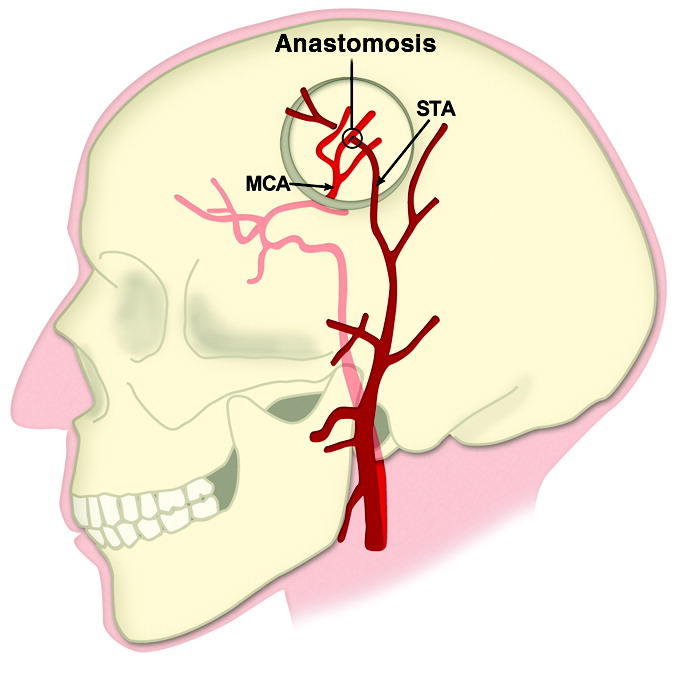 STA-MCA bypass — microsurgical anastomosis under the microscope
What it is: The Superficial Temporal Artery to Middle Cerebral Artery (STA-MCA) bypass is a microsurgical procedure that creates a new blood supply route to ischaemic (oxygen-starved) brain tissue — bypassing a blocked or narrowed internal carotid or middle cerebral artery.
When it is needed: Moyamoya disease, complex intracranial aneurysms requiring parent artery sacrifice, carotid artery occlusion with haemodynamic compromise, and selected patients with recurrent TIAs or strokes despite maximal medical therapy.
The procedure: Under a microscope, the superficial temporal artery (STA) — a small scalp artery — is anastomosed (connected) end-to-side to a branch of the MCA on the brain’s surface through a small craniotomy. Blood flow is immediately re-established to the under-perfused territory, reducing future stroke risk.
Outcomes: In carefully selected patients, STA-MCA bypass dramatically reduces the risk of recurrent stroke and improves cerebral perfusion. At our centre, intraoperative indocyanine green (ICG) videoangiography confirms graft patency immediately after anastomosis.
Haematoma EvacuationMinimally Invasive Haematoma Evacuation
 Minimally invasive endoscopic haematoma evacuation
What it is: Haemorrhagic stroke (bleeding in the brain) creates a haematoma that compresses surrounding brain tissue. Surgical evacuation removes the clot, relieves pressure, and stops further neurological deterioration.
Minimally invasive approach: Rather than large open craniotomies, we use stereotactic-guided and endoscopic techniques to evacuate intracerebral haematomas through the smallest possible opening — reducing surgical trauma, blood loss, and recovery time while achieving effective clot removal.
Indications: Large lobar haematomas, cerebellar haematomas causing brainstem compression, haematomas with deteriorating neurological status, and intraventricular haemorrhage with hydrocephalus.
Neuronavigation guidance: Every haematoma evacuation is planned with neuronavigation — ensuring the most direct, least damaging route to the clot.
Carotid EndarterectomyCarotid Endarterectomy (CEA)
 Carotid endarterectomy — surgical plaque removal
What it is: Carotid endarterectomy surgically removes atherosclerotic plaque from the carotid artery in the neck — the artery supplying blood to the brain. Severe carotid stenosis is a major cause of ischaemic stroke and TIA.
The procedure: Through a neck incision, the carotid artery is opened and the plaque is meticulously removed, restoring full blood flow to the brain. The artery is then closed with a patch graft to prevent re-narrowing. The procedure is performed under intraoperative neuromonitoring (SSEP and EEG) to detect any brain ischaemia during the carotid clamp period.
Who benefits: Symptomatic carotid stenosis >50% (patients who have had a TIA or minor stroke) and asymptomatic stenosis >70%. CEA reduces the risk of a major stroke by up to 50–70% in symptomatic patients.
Our approach: CEA is performed at our centre by a neurosurgeon with dedicated vascular training, with all patients reviewed in a multidisciplinary stroke team including neurology, neuroradiology, and cardiology.
ThrombectomyMechanical Thrombectomy for Acute Ischaemic Stroke
 Mechanical thrombectomy — stent retriever restoring blood flow within minutes
In large vessel occlusion stroke, mechanical thrombectomy physically removes the clot from the blocked cerebral artery — achieving immediate reperfusion of the ischaemic territory. This procedure must be performed within 6–24 hours of stroke onset and has revolutionised outcomes for major ischaemic stroke. A stent retriever or aspiration catheter captures and removes the clot under fluoroscopic guidance.
Vascular NeurosurgeryVascular Diseases of the Brain & Spine
Abnormalities of the brain’s blood vessels — aneurysms, AVMs, cavernomas, carotid stenosis — carry a risk of catastrophic haemorrhage or stroke. We offer both open microsurgical and endovascular management.
Advanced Neuro-Intervention DepartmentOne of India’s Most Advanced Cerebrovascular Centres
At Fortis Hospital, Manesar, we have access to one of India’s most advanced neuro-intervention departments — combining cutting-edge endovascular technology with expert open microsurgery. For each patient, the best approach — surgical, endovascular, or combined — is chosen based on anatomy, lesion characteristics, and patient factors. Our team has extensive experience with coiling, flow diverters, embolisation, stenting, and open clipping — performing the full spectrum under one roof.
AneurysmIntracranial Aneurysm
 Intracranial aneurysm
An aneurysm is a balloon-like bulge in a brain artery. Rupture causes subarachnoid haemorrhage — one of the most devastating neurological events. Management options: Microsurgical clipping (placing a metal clip across the aneurysm neck through a craniotomy) or endovascular coiling (filling the aneurysm sac with platinum coils via a catheter in the groin). The choice depends on aneurysm shape, location, neck geometry, and patient factors. Flow diverters (Pipeline Embolisation Device) are used for wide-neck or fusiform aneurysms. All ruptured aneurysms are managed as emergencies.
AVMArteriovenous Malformation (AVM)
 Arteriovenous malformation of the brain
An AVM is an abnormal tangle of blood vessels short-circuiting the normal capillary bed. It may bleed, cause seizures, or produce progressive neurological deficits. Management: Microsurgical resection for accessible AVMs, pre-operative embolisation to reduce blood flow before surgery, stereotactic radiosurgery for small deep AVMs, or a combined multimodal approach. The Spetzler-Martin grading system guides treatment decisions. At our centre, complex high-grade AVMs are managed with combined embolisation and surgery under neuronavigation guidance.
Vascular MeningiomaVascular Meningiomas
 Vascular meningioma — pre-op embolisation reduces surgical bleeding
Some meningiomas have an exceptionally rich blood supply, making surgery high-risk for major intraoperative haemorrhage. At our centre, we routinely perform pre-operative embolisation of these tumours — a neuro-interventional procedure that occludes the tumour’s blood supply 24–48 hours before open surgery. This dramatically reduces intraoperative bleeding, shortens operating time, and improves the safety of complete resection for even the most vascular meningiomas.
Carotid StenosisCarotid Artery Stenosis & Carotid Artery Stenting (CAS)
 Carotid artery stenting — restoring blood flow to the brain
Narrowing of the carotid artery by atherosclerotic plaque. When surgical endarterectomy (CEA) is not preferred — due to anatomical factors, radiation-induced stenosis, or medical comorbidities — carotid artery stenting (CAS) is performed via catheter through the groin. A stent is deployed across the plaque to keep the artery open, with a distal embolic protection device to catch any dislodged debris. Results are comparable to CEA in appropriately selected patients.
CavernomaCavernous Malformations (Cavernoma)
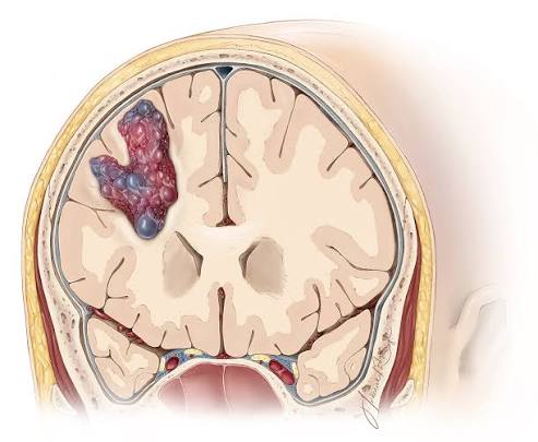 Cavernoma on MRI
Small vascular lesions that bleed repeatedly, causing progressive neurological deficits or epilepsy. Microsurgical removal under intraoperative neuromonitoring is highly effective — the excellent plane between cavernoma and surrounding brain allows complete removal with minimal brain retraction.
Endovascular ProceduresNeuro-Interventional Procedures
Modern cerebrovascular disease management often does not require open brain surgery. Catheter-based endovascular techniques treat aneurysms, malformations, stenoses, and strokes through small punctures in the groin — with no craniotomy required.
State-of-the-Art Neuro-InterventionWorld-Class Endovascular Technology
Our neuro-intervention department at Fortis is equipped with a biplane digital subtraction angiography (DSA) suite and the full armamentarium of modern endovascular devices. Our interventional team performs all procedures in a dedicated neuro-intervention suite with 24/7 emergency availability for acute stroke thrombectomy and ruptured aneurysm coiling.
CoilingEndovascular Coiling of Aneurysms
 Coiling — platinum microcoils filling the aneurysm sac
Under general anaesthesia, a microcatheter is navigated through the femoral artery in the groin, through the aorta, and into the cerebral circulation — reaching the aneurysm. Platinum microcoils are deployed to fill the aneurysm sac, causing it to clot and seal off from the circulation. Patients are typically mobile the next day and discharged within 2–3 days. No head shaving, no craniotomy, no scar.
Flow DiverterFlow Diverter (Pipeline Embolisation Device — PED)
 Pipeline embolisation device — flow diverter for complex aneurysms
A braided, high-mesh-density stent deployed across the neck of a complex or large aneurysm. The PED redirects blood flow away from the aneurysm, causing it to gradually thrombose and shrink over weeks to months. It is particularly effective for large, wide-neck, or fusiform aneurysms of the internal carotid artery not suitable for conventional coiling.
EmbolisationAVM & Tumour Embolisation
Pre-operative embolisation of AVMs reduces their blood supply before open surgical resection — making surgery safer, reducing blood loss, and increasing the chance of complete removal. Similarly, highly vascular tumours (meningiomas, paragangliomas, juvenile nasopharyngeal angiofibromas) are embolised before surgery to minimise haemorrhage.
StentingIntracranial Stenting & Carotid Stenting
Carotid artery stenting with embolic protection
Stents are deployed in narrowed intracranial or carotid arteries to restore blood flow. Intracranial angioplasty and stenting is used for severe symptomatic intracranial atherosclerosis not responding to medical treatment. Carotid artery stenting with embolic protection is offered as an alternative to open carotid endarterectomy in selected patients.
ThrombectomyMechanical Thrombectomy for Acute Ischaemic Stroke
Mechanical thrombectomy — clot retrieval for acute ischaemic stroke
In large vessel occlusion stroke, mechanical thrombectomy physically removes the clot from the blocked cerebral artery — achieving immediate reperfusion of the ischaemic territory. This procedure must be performed within 6–24 hours of stroke onset and has revolutionised outcomes for major ischaemic stroke. A stent retriever or aspiration catheter is used to capture and remove the clot.